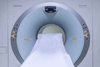
Diffusion MRI Imaging: What You Need to Know
Diffusion MRI, or magnetic
resonance imaging, creates in vivo pictures of biological tissues that are
weighted by local microstructural water diffusion features. Each picture voxel
has an image intensity that reflects a single best assessment of the rate of
water diffusion at that place in diffusion-weighted imaging. Other MRI
measurement methods, such as T1 or T2 relaxation rates in MRI cost Miami, are less sensitive to early changes following a
stroke than this assessment.
In diffusion MRI imaging, a high
signal-to-noise ratio is critical (DWI). This is usually accomplished by
improving gradient performance to lower the time necessary for diffusion
sensitization and signal capture, hence reducing the DWI sequence's minimum
feasible time-to-echo (TE). Parallel imaging can also be utilized to shorten
the amount of time it takes to acquire a signal.
Image quality is improved further
by higher gradient performance and parallel imaging, which reduce distortion
and susceptibility artefacts.
Higher gradient performance is
beneficial to DWI applications, as DWI sequences typically demand the full
capability of gradient performance. However, due of safety restrictions based
on physiological stimulation limits, gradient systems are only likely to
improve marginally in the future compared to the high-specification MRI systems
already available.
Any approach to actively limit the
effect of eddy current-induced spatial distortions, such as specific diffusion
encoding techniques or optimized gradient coil design, would also benefit
overall image quality. Actions made during the capture phase are often
preferable than post-processing procedures.
DWI experiments using an MRI
system with a stronger magnetic field would be an alternative technique
to increase the SNR of the DW image. For example, from a 1.5T system to a 3T
system, the SNR is projected to increase by double. As a result, DWI studies on
3T systems can benefit from a wider image contrast range than analogous 1.5T
studies. Image quality becomes more vulnerable to susceptibility and distortion
artefacts as the magnetic field increases.
As a result, greater gradient
specifications and parallel imaging are more relevant in DWI studies on 3T RI
systems, and give significant picture quality improvements.
Diffusion Fat tissues can cause
signal misregistration in MRI imaging. Special fat-suppression techniques are
utilized to reduce the fat signal, and their success is likely to be reliant on
the primary magnetic field homogeneity. DWI is more susceptible to hardware
flaws than traditional imaging applications. Image quality degradation in DWI
is frequently linked to issues with the performance of gradient or shimming coils.
If at all practicable, DWI should be included in a quality-control procedure,
particularly in long-term, comparative, or quantitative investigations.
Diffusion MRI imaging sequences
last only a few seconds, thus they can be utilized as a supplement to
traditional MRI studies without adding too much to the scan time. Because the
b-value is the primary determinant of image contrast, DWI sequences are also
straightforward to execute. Furthermore, no contrast agent infusion or
physiological monitoring is required. Electrocardiogram gating, on the other
hand, can help with sophisticated DWI analysis, such as DTI computation.
Although the hazards are minimal in this mode, and scanning with DWI is
incredibly quick, there is a slight chance of mild peripheral nerve
stimulation.
Furthermore, because acoustic noise
levels in DWI sequences are higher than in conventional MRI, hearing protection
should be supplied to reduce patient discomfort and prevent temporary hearing
loss.
Visit For More Info:

Comments
Post a Comment